2026-06-20
Lateral Canthoplasty Revision: Surgical Methods and Before & After Photos
Learn about lateral canthoplasty revision, common side effects like U-shaped eye corners, and the surgical process with before and after results from Gangnam Seoyeon Plastic Surgery.
Hello!
At Gangnam Seoyeon Plastic Surgery, we always strive to provide the highest level of patient satisfaction :)
Today, we will take a look at lateral canthoplasty revision performed by Dr. Choi Dong-il at Gangnam Seoyeon Plastic Surgery.
We will explain who needs this procedure, the surgical methods involved, and share some before and after photos.
The photos below have not been edited in any way, except for brightness adjustments :)

Lateral Canthoplasty Side Effect 1 - ‘U-shaped’ Eye Corners
When lateral canthoplasty is performed excessively, various side effects can occur.
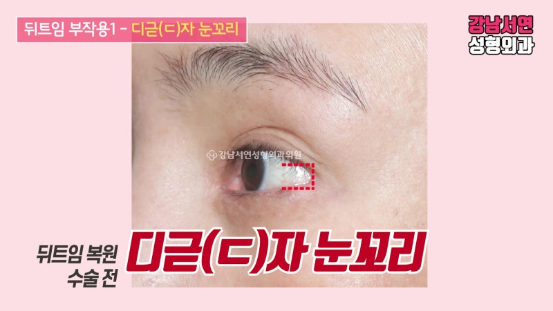
The first common side effect is the ‘U-shaped’ (ㄷ-shaped) eye corner.
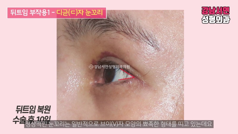
While it varies by individual, a normal eye corner typically has a sharp, ‘V-shaped’ appearance.
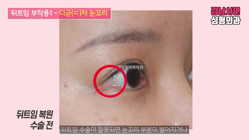
If the surgery is unsuccessful, the corner of the eye may gap open,
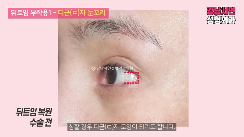
and in severe cases, it takes on a blunt ‘U-shape’ (ㄷ-shape).
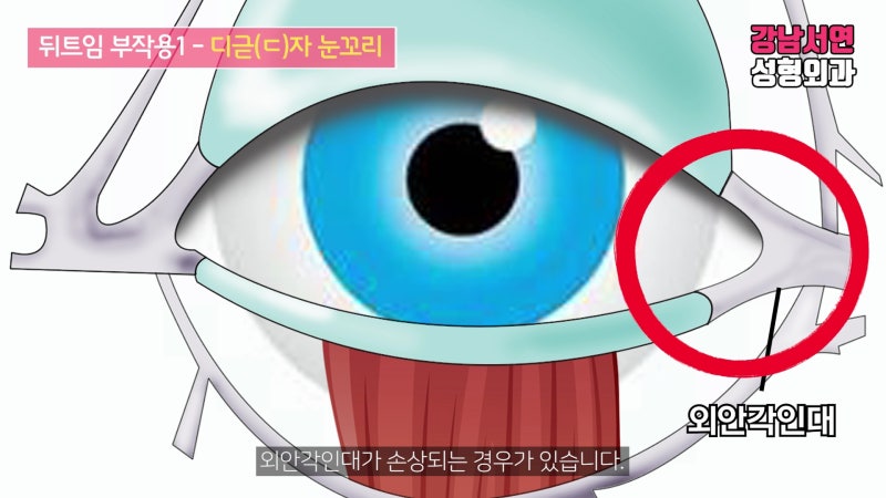
The reason this happens is often due to damage to the lateral canthal tendon during an overly aggressive procedure.
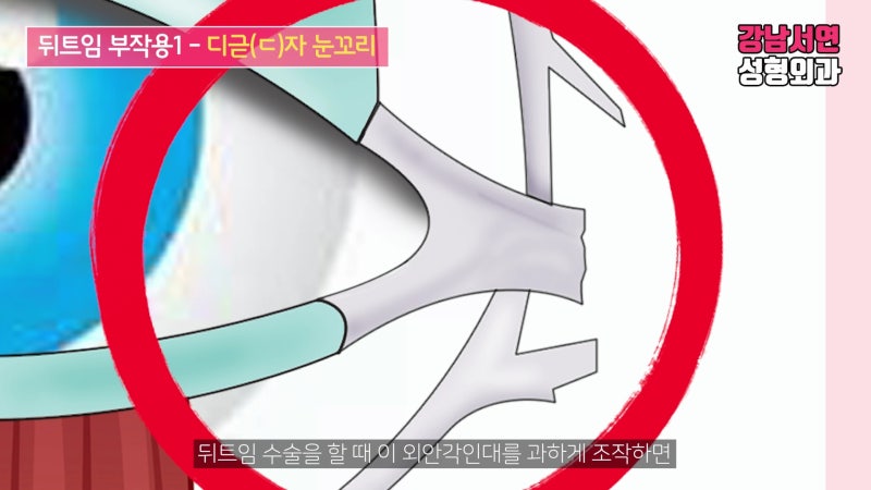
The lateral canthal tendon is attached to the lower eyelid and supports the eye from both sides, helping it close properly.
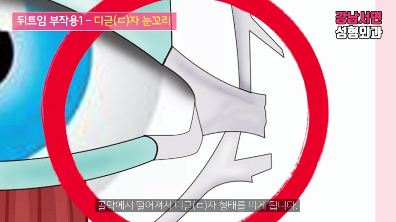
If this tendon is manipulated excessively during surgery, it can become detached from the periosteum, resulting in that ‘U-shaped’ appearance.
Lateral Canthoplasty Side Effect 2 - Mucosal and Conjunctival Exposure
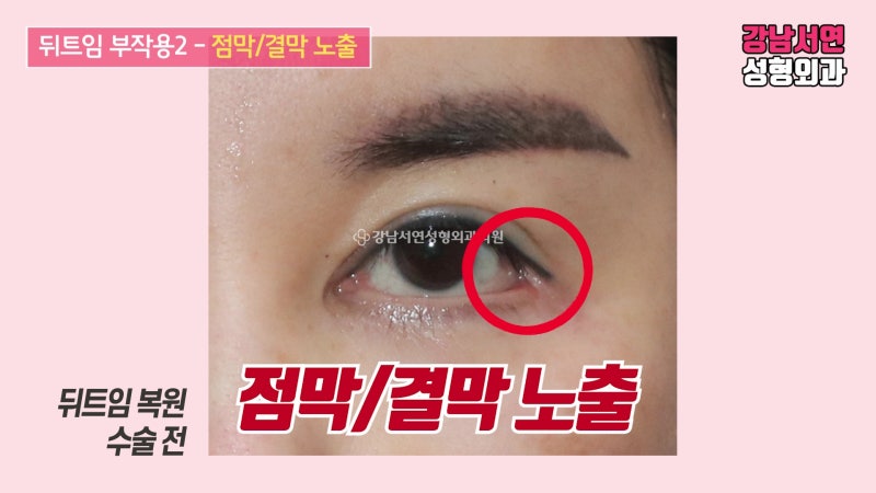
The second side effect is the exposure of the mucosa or conjunctiva after surgery.
This is also related to the lateral canthal tendon.
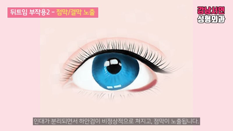
When the lateral canthal tendon is damaged due to excessive surgery, the tendon separates, causing the lower eyelid to sag abnormally and exposing the red mucosa.
In severe cases, the eye may not close fully, and the patient may appear to have a ‘sad’ or drooping expression.
Lateral Canthoplasty Revision Method
So, how is lateral canthoplasty revision performed?
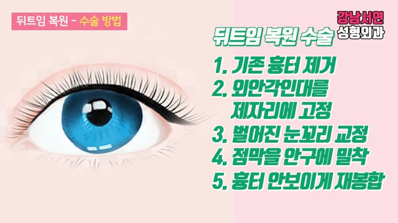
The revision process involves:
1. Cleanly removing the existing scar tissue.
2. Locating the detached lateral canthal tendon and restoring it to its original position.
3. Reshaping the gapped eye corner back to a natural form.
4. Re-attaching the sagging or everted mucosa closely to the eyeball.
5. Re-suturing while minimizing the visibility of the scar.
Revision Before & After Photos
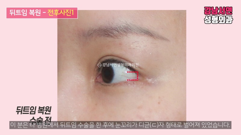
Let’s look at some before and after photos.
This patient had a lateral canthoplasty at another clinic, which left the eye corners gapped in a U-shape.
The key to this revision was removing the old scar tissue and securing the detached tendon back to the periosteum to prevent recurrence while hiding the incision line.
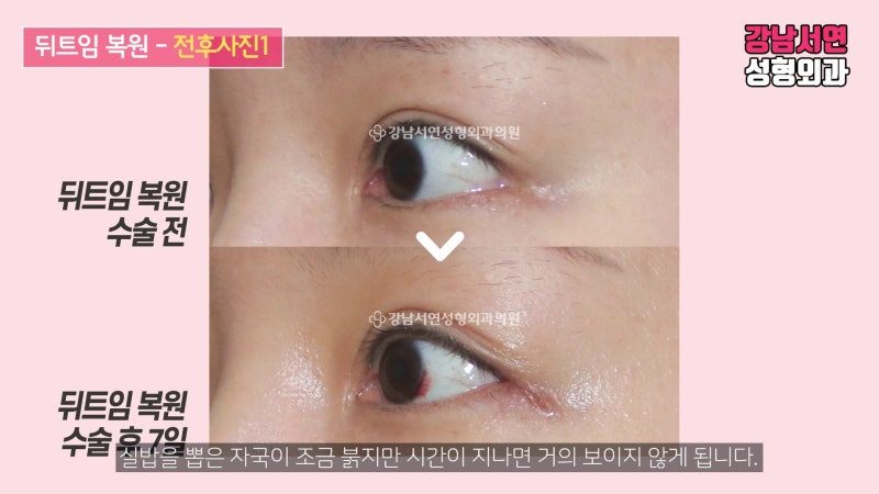
This is the appearance one week after surgery.
The stitch marks are slightly red, but this will fade and become almost invisible over time.
Here is another case.
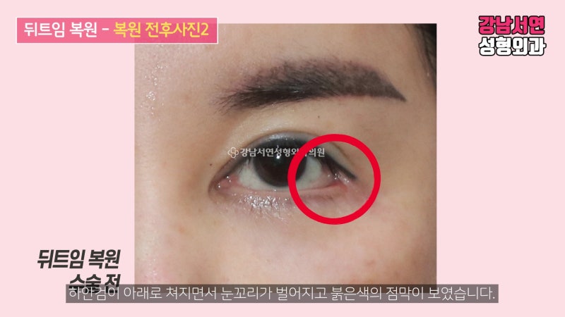
After a previous surgery elsewhere, this patient’s lower eyelid sagged, the corner gapped, and the red mucosa was visible.
The lower eyelid looked like a ‘sagging clothesline’.
In this case, you can think of the revision as pulling that sagging clothesline taut again.
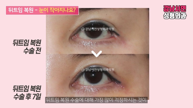
This is the appearance one week after surgery.
You can find more detailed information about lateral canthoplasty revision in the video below.
↓↓↓↓↓↓
Gangnam Seoyeon Plastic Surgery complies with medical laws. The photos in this post were taken under identical conditions with the patient’s prior consent. Please note that side effects such as bleeding, infection, and inflammation may occur after plastic surgery or procedures depending on the individual.
- Single-specialist system led by the Representative Director - Accurate diagnosis of eye condition / recommending only necessary surgeries - Operating rooms equipped with CCTV |
Feel free to visit us for a 1:1 consultation with our plastic surgery specialist :)
If you have any questions or would like a consultation, please leave a comment, contact us via KakaoTalk Plus Friend @GangnamSeoyeonPlasticSurgery, or call us at 02-535-8889 :)






#GangnamSeoyeonPlasticSurgery #GangnamSeoyeon #SeoyeonPlasticSurgery #LateralCanthoplasty #LateralCanthoplastyRevision #EyeSurgerySideEffects #EyelidSurgery #BeforeAndAfter #DrChoiDongIl
Frequently Asked Questions
Why does the outer corner of the eye become D-shaped after lateral canthoplasty?
This is due to damage to the lateral canthal ligament from excessive surgery. If the lateral canthal ligament, which supports the eye during canthoplasty, is manipulated too much, it can detach from the periosteum, leading to a D-shaped outer corner of the eye instead of the normal V-shape.
Can the conjunctiva be exposed as a side effect of lateral canthoplasty?
Yes, if the lateral canthal ligament is damaged, the mucous membrane or conjunctiva can be exposed. As the ligament separates, the lower eyelid sags abnormally, and the red mucous membrane becomes visible. In severe cases, the eye may not close completely, or it may appear as if one is constantly crying.
What is the process for lateral canthoplasty revision surgery?
The process involves removing existing scars and returning the damaged ligament to its original position. The detached lateral canthal ligament is located and fixed to the periosteum to restore the outer corner of the eye to its normal shape, the sagging mucous membrane is reattached to the eyeball, and the area is meticulously re-sutured to minimize visible scarring.
When are the stitches removed after lateral canthoplasty revision surgery?
The stitches are removed one week after the surgery. Immediately after stitch removal, the area may appear slightly red, but over time, the redness will subside, and the scar will naturally heal to be almost invisible.